Search my links!

Showing posts with label ultrasound. Show all posts

Sunday, 17 April 2011

Carotid US

Anatomy:


- Circle of Willis is supplied by the carotids (anterior circulation) + vertebrals (posterior circulation).
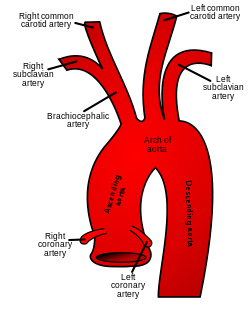
- On the RIGHT the CCA originates from the BRACHIOCEPHALIC TRUNK.
- On the LEFT the CCA originates from the AORTIC ARCH directly.
Gray506.svg
- The points of origin are infrequent points for plaque formation.
- Stenosis most frequently occures at the level of bifurcation of the CCA into ICA and ECA.
- The first intracranial branch of the ICA is the ophtalmic artery.
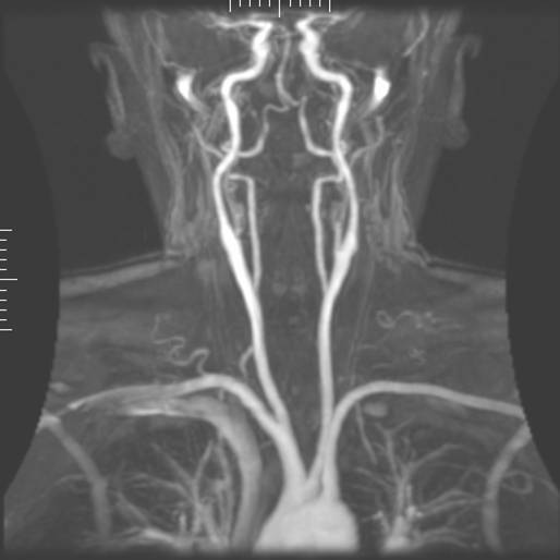
- The vertebral artery (supplying the post. circulation of the circle of Willis) usually arises from the subclavian artery, see below:


- The vertebral artery is divided into 5 segments ( not important for us in examining the carotids).
- The vertebral artery maybe hypoplastic in certain segments, and its usually more prominent on one side.
- The vertebral arteries unite to form the barilar arteries -> circle of Willis -> posterior cerebral artery.
File:Mra1.jpg

What is subclavian steal syndrome?

In Subclavian steal syndrome (SSS) a reduced quantity of blood flows through the proximal subclavian artery. As a result, blood travels up one of the other blood vessels to the brain (the other vertebral or the carotids), reaches the basilar artery or goes around the cerebral arterial circle and descends via the (ipsilateral) vertebral artery to the subclavian (with the proximal blockage) and feeds blood to the distal subclavian artery (which supplies the upper limb and shoulder).
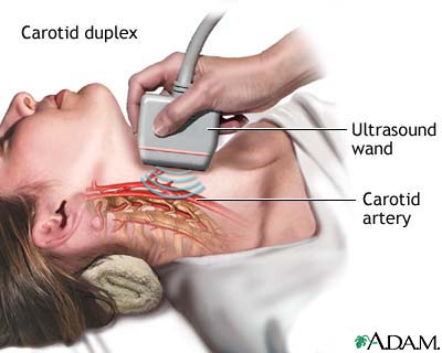

Scaning the carotids:

  • normal
    • ICA PSV is less than 125 cm/sec and no plaque or intimal thickening is visible sonographically.
    • additional criteria include ICA / CCA PSV ratio < 2.0 and ICA EDV < 40 cm/sec.
  • < 50% ICA stenosis
    • ICA PSV is less than 125 cm/sec and plaque or intimal thickening is visible sonographically.
    • additional criteria include ICA / CCA PSV ratio < 2.0 and ICA EDV < 40 cm/sec.
  • 50 - 69% ICA stenosis
    • ICA PSV is 125 - 230 cm/sec and plaque is visible sonographically.
    • additional criteria include ICA / CCA PSV ratio of 2.0 - 4.0 and ICA EDV of 40 - 100 cm/sec.
  • > / = 70% ICA stenosis but less than near occlusion
    • ICA PSV is greater than 230 cm / sec and visible plaque and luminal narrowing are seen at gray-scale and color Doppler US (the higher the Doppler parameters lie above the threshold of 230 cm/sec, the greater the likelihood of severe disease).
    • additional criteria include ICA / CCA PSV ratio > 4 and ICA EDV > 100 cm/sec.
  • near occlusion of the ICA
    • velocity parameters may not apply, since velocities may be high, low, or undetectable.
    • diagnosis is established primarily by demonstrating a markedly narrowed lumen at colour or power Doppler US (35).
  • total occlusion of the ICA
    • no detectable patent lumen at gray-scale US and no flow with spectral, power, and colour Doppler US.

    -----------------------------------------------------------------------------------------------------------------------------------------------------------------------

    Carotid arterial disease can be conveniently graded into percentage stenoses by assesing several sonographic parameters. The NASCET criteria is as follows.

    • no stenosis : normal wave form
    • < 15 % stenosis :
      • deceleration spectral broadening with a peak systolic velocity (PSV) of < 125cm/s
    • 16 - 49 % stenosis :
      • pan-systolic spectral broadening with a peak systolic velocity (PSV) of < 125 cm/s
    • 50 - 69 % stenosis :
      • pan-systolic spectral broadening with a peak systolic velocity (PSV) of > 125 cm/s and
      • end diastolic velocity (EDV) < 110 cm/s or
      • ICA/CCA PSV ratio > 2 but < 4
    • 70 - 79 % stenosis :
      • pan-systolic spectral broadening with PSV > 270 cm/s or
      • EDV > 110 cm/s or
      • ICA/CCA PSV ratio > 4
    • 80 - 99% stenosis : EDV > 140 cm/s
    • complete occlusion : no flow : terminal thump
    ------------------------------------------------------------------------------------------------------------------------------------------------------------------------


    --------------------------------------------------------------------------------------------------------------------------------------------------------------

    LINKS:

    Video:
    http://www.youtube.com/watch?v=yuKu_6RhNOM

    (Text source: Teaching atlas of CDS-Thieme, wikipedia)
    (Images: google pictures)

    Tuesday, 12 April 2011

    Renal arteries in general. The transplanted kidney


    TRANSPLANT KIDNEY


    Vascular supply from end-to-side
    Anastomosis of donor artery and vein to external iliac artery and vein. If multiple arteries, usually joined with single anastomosis to EIA. Ureter anastomosed to superolateral wall of urinary bladder. En bloc transplant of a set or pediatric kidneys with caudal ends of IVC and Aorta anastomosed end-to-side to recipient's EIA and EIV.


    Usually parallel to incision with hilum inferiorly and posteriorly, extra-peritoneal location. Obtain longitudinal and transverse measurements. Usually hypertrophies ~ 15% in first 2 weeks and may increase by 40% in first 6 months.

    Because kidney is more superficial, pyramids are more easily visualized, accentuating the cortico-medullary differentiation. Evaluate for any intrinsic pathology: calculi, tumors, etc. Collections, hydronephrosis.


    Post- operative hematomas- appearance depends upon chronicity. Urine leaks or urinomas- due to anastomotic leaks or ureteric ischemia. Appear well defined, anechoic with occasional hydronephrosis. May use radionuclide imaging to confirm nature of collection. Lymphoceles- occur 4 – 8 weeks after surgery in 15%. May obstruct ureter or veins. Appear well defined and either anechoic or with fine septations.


    Screen and image with color/power, looking for focal and/or diffuse hypoperfusion. Obtain spectral traces of interlobar arteries in upper, mid and lower poles with appropriate factors. Image and obtain spectral Doppler of main renal artery and vein and EIA and EIV.


    Arteries- brisk upstroke, low resistance with normal RI of 0.6 to 0.8. Normal velocity main renal artery <200 cm/sec. Veins- may be monophasic with continuous flow or demonstrate some pulsatility with cardiac cycle.


    ATN:
     Occurs to some extent in all cadaveric transplants. Most common cause of delayed graft function (need for dialysis in 2 weeks post transplant). Non-specific imaging features: normal or changes in echogenicity, qualitatively decreased color flow, RI may be normal or increased.


    Rejection:
    Hyperacute- rarely imaged since it occurs during surgery. Acute- occurs in up to 40% in first few weeks and is a poor long term prognostic indicator. Similar US and radionuclide findings. US findings non-specific.

    Most common cause of late graft loss. Progressive loss renal function beginning 3 months after transplant. Patients with acute rejection are predisposed. US- Cortical thinning, mild hydronephrosis, prominent sinus fat, dystrophic calcifications, decreased color, normal or increased RI.


    Hydronephrosis
    Obstruction is rare, though will usually be at UV junction from stricture or intra-luminal lesion. Mild pelvocaliectasis may be 2º overhydration, decreased ureteric tone, U-V reflux.


    Renal artery thrombosis
    Occurs in < 1% typically within first month. Most common cause is acute rejection with retrograde thrombosis of small to large arteries. Other causes: pediatric kidneys, emboli, acquired stenosis, hypotension, vascular kink, hypercoagulable state, poor anastomosis, trauma. US: Absent arterial and venous flow in kidney and main renal artery.


    Most common vascular complication in up to 10% in first year. Three possible sites:
     – Donor portion, typically at end-to-side anastomosis
    – Recipient portion- more uncommon, from intraoperative clamp or intrinsic atherosclerosis
    – At anastomosis- more frequent in end-to-end anastomoses

    US Findings of RAS
     Parvus-tardus flow in intra-parenchymal vessels. Use color Doppler to locate stenosis. Peak systolic velocities > 200 cm/sec with turbulent flow. False positive diagnoses may occur with abrupt turn in the main renal artery. With chronic rejection, segmental renal artery stenoses may occur.


    AVM and PSA
     Both are typically the result of trauma during percutaneous biopsy. AVM- color shows focal area of mixed colors occasionally with feeding vessels. AVM produces vibrations which result in color assigned to the perivascular tissues.
    PSA- may appear as a simple cyst on grey scale imaging but with typical swirling arterial flow on Doppler.


    RV thrombosis
    Occurs in up to 4% transplants. Sx: acute pain 2° swelling of kidney, oliguria in first week. Causes: surgical difficulties, hypovolemia, femoral/iliac vein thrombosis, compression by collections. US- absent venous flow, reversal of diastolic flow in artery. Kidney may be enlarged, hypoechoic.


    RV stenosis
    Grey Scale- normal or hypoechoic. Color Doppler- aliasing at stenotic site. Spectral Doppler- 3 to 4 times increase in velocity across the region indicates a hemodynamically significant stenosis.