Search my links!

Tuesday 10 May 2011

Orthopaedics

Legg-Calvé-Perthes disease:


- is idiopathic avascular necrosis (AVN) of the growing femoral epiphysis seen in children, and should not be confused with the Perthes lesion of the shoulder.

- ages of 4 - 8 years, and the most frequent presenting feature is pain with or without a limp. In 10 - 12 % of cases both hips are involved.

Plain film

Early signs
  • asymmetrical femoral epiphyseal size (smaller on affected side)
  • apparent increased density of the femoral head epiphysis
  • widening of the medial joint space
  • blurring of the physeal plate (stage 1: see staging of Legg-Calve-Perthes syndrome)
  • radiolucency of the proximal metaphysis
Late signs

Eventually, the femoral head begins to fragment (stage 2), with subchondral lucency (crescent sign) and redistribution of weight-bearing stresses leading to thickening of some trabeculae which become more prominent.

The typical findings of advanced burnt out (stage 4) Perthes disease are:

  • femoral head deformity with widening and flattening
  • proximal femoral neck deformity

Additionally, tongues of cartilage sometimes extend inferolaterally into the the femoral neck, creating lucencies, which must be distinguished from infection or neoplastic lesions. The presence of metaphyseal involvement no only increases the likelihood of femoral neck deformity, but also make early physeal closure with result leg length disparity more likely.


(Source : www.radiopedia.org)

Monday 9 May 2011

Contrast enhanced exams of the GIT

COLON TRANSIT TEST / or TIME

The simplest and most easily interpreted colonic transit evaluation is performed with a plain abdominal radiograph on day 5 after the patient has ingested a gelatin capsule containing radiopaque markers (SITZMARKS™, Konsyl Pharmaceuticals, Fort Worth, TX). Each capsule contains 24 radiopaque polyvinyl chloride o-rings of 1 mm x 4.5 mm. The test is inexpensive and well tolerated by patient.


Indication
Adult patients with severe constipation but otherwise negative G.I. evaluations

Procedure
On day 0, direct the patient to take one SITZMARKS capsule by mouth with water. Instruct the patient to use no laxatives, enemas, or suppositories for 5 days.

On day 5, obtain a plain abdominal radiograph (KUB). Determine the number and location of remaining radiopaque rings.

Patients who expel at least 80% of the markers by 5 days (5 or fewer rings remaining) have grossly normal colonic transit.

For patients who retain 6 or more markers, one may elect to get a follow-up radiograph on day 7, when normally 100% of marker should have been expelled.


Interpretation of Results

If over 80% of the radiopaque markers (19 or more) are passed by day 5, colonic transit is grossly normal.

If 6 or more of the markers remain on day 5, this is abnormal:

If remaining markers are scattered about the colon, the problem is most likely colonic hypomotility or inertia.

If remaining markers are accumulated in the rectum or rectosigmoid, the condition is most likely functional outlet obstruction, such as rectal intussusception or anismus


    1. Figure 1

Its a useful in evaluating patients with constipation, abdominal bloating, and refractory irritable bowel syndrome.
Colonic transit time has been traditionally measured using radiopaque markers.
Several methods have been suggested, including the single-marker bolus technique (ingestion of markers on a specific day followed by several x-rays until all markers are passed) or multiple-marker bolus technique (ingestion of markers each day for several days followed by a limited number or a single abdominal x-ray).

This test has a relatively long duration (5-7 days) and requires the patient to abstain from laxatives, enemas, and other medications known to affect gastrointestinal motility. These drawbacks have led to a desire to develop alternatives to this study.

Colonic scintigraphy can be used as an alternative to the radiopaque marker technique for measuring colonic transit time. This test involves administration of radioactive isotope and following its progression through the gastrointestinal tract with a large-field-view gamma camera. The test uses long half-life radionuclides indium 111-diethylenetriamine pentaacetic acid (111In-DTPA) or iodine 131.

LINKS & Sources:

CT scans


Intraventricular haemorrhage:

Most commonly seen in nfants, but never the less can be seen in adults also.
There are two main ways in which IVH can potentially cause damage. First, IVH may affect the flow of CSF in the ventricles and second, IVH may cause damage to brain tissue adjacent to the ventricles. Once damage has occurred to brain tissue, it cannot be reversed.

  • Grade 1 - bleeding occurs just in a small area of the ventricles.
  • Grade 2 - bleeding also occurs inside the ventricles.
  • Grade 3 - ventricles are enlarged by the blood.
  • Grade 4 - bleeding into the brain tissues around the ventricles.

Read more: http://www.articlesbase.com/diseases-and-conditions-articles/intraventricular-haemorrhage-ivh-2057162.html#ixzz1SjdeaTg4
Under Creative Commons License: Attribution No Derivatives


Links:
http://en.wikipedia.org/wiki/Intraventricular_hemorrhage


------------------------------------------------------------------------------------------------


Initially thought to be a thrombus, later on proven to be a Myxoma found at the base of the pulmonary trunk.

Complete resection in typical cases is usually curative.

Myxomas, as is the case with other cardiac tumours, appear as intra-cardiac masses, most often in the left atrium and attached to the interatrial septum. They are usually heterogeneously low attenuating ( approximately 2/3 of cases ). Due to repeated episodes of haemorrhage, dystrophic calcification is common .

Links:
-------------------------------------------------------------------------------------------------


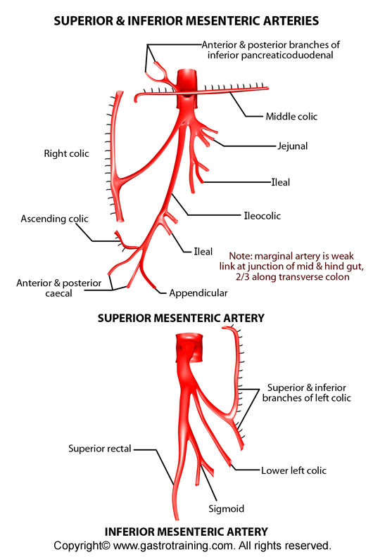
SMA occlusion

Acute occlusion can be due to a number of causes:

  • embolic event : ~ 60%
  • acute in situ thrombosis superimposed on atherosclerosis : 30%
  • aortic dissection with involvement of the SMA origin
  • slow flow / idiopathic
CT findings:

  • lack of enhancement of the lumen of the SMA and / or its branches
    • embolism lodgement location varies
      • 15% origin
      • 50% just distal to the origin of the middle colic artery
  • bowel wall thickening
  • ileus
  • absent mucosal enhancement
  • free fluid
  • mural gas, portal venous gas or free intra abdominal gas

LINKS:

------------------------------------------------------------------------------------------------


Elongated ICA / Kinking of the ICA


Tortuosity of the cervical ICA is not a rare condition and they can easily be mistaken clinically for an aneurysm, a tumor or an abscess and subsequently injured during an attempted biopsy or excision. Thus, regardless the controversy of its causes (congenital or acquired) it should be included in the differential diagnosis of cervical soft tissue widening. Also, they should be taken into consideration on the diagnostic procedures for ischemic transitory attacks and/or stroke.

Links:

-------------------------------------------------------------------------------------------------
Intracardiac tumor +/- thrombus

Links:
http://www.med-ed.virginia.edu/courses/rad/cardiacmr/Techniques/Contrast.html


-------------------------------------------------------------------------------------------------


Tumor in the pylorus

Links: